Skip to content
Navigate to homepage - Cerba Research

Multiplex IHC & Immunofluorescence (IF)

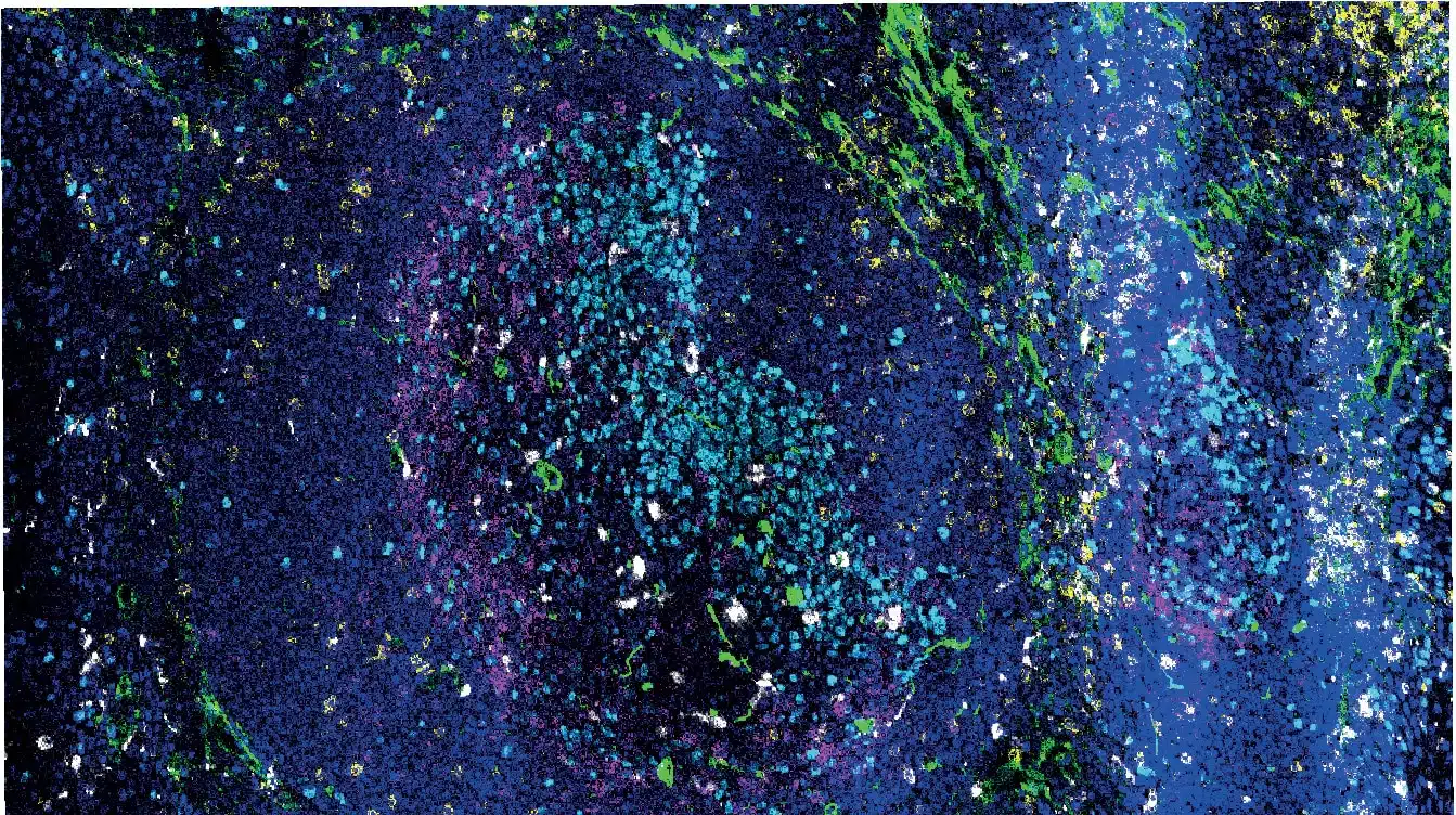
Multiplex IHC & Immunofluorescence (IF) offers a technical advantage by allowing the detection of co-expression and spatial organization of up to 8 targets within a preserved tissue architecture, on a single section.

Your Project Deserves Our Expert Multiplex IHC Team

Cerba Research was one of the first organizations in Europe to offer multispectral multiplex IHC services to pharmaceutical and biotechnology companies.

​​In an era where increased numbers of protein biomarkers, advanced phenotyping, and spatial context are needed on precious samples of limited size, multiplex IHC is the right approach to obtain the most relevant and comprehensive data from tissue samples.

Our multiplex IHC panels are the right tool to understand the sociology (distances between cell populations, tissue infiltration, and advanced phenotyping based on both the expression or absence of expression of biomarkers) of key cell players and biomarkers in the tissue.

With years of experience testing reagents and technical solutions for multiplex IHC (including Akoya, Roche, Ultivue, Cell IDx, and Enzo Life Sciences), our science team has the expertise to propose the most scientifically relevant and prone-to-success solution to every program.

Cerba Research provides standard workflows for the development of chromogenic (up to 3 colors) or immunofluorescence (up to 8 colors) multiplex IHC panels, as well as virtual multiplexing.

Multiplex IHC panels development always begins with the initial chromogenic simplex protocols and keeps referencing back to the chromogenic IHC gold standard throughout the multiplex optimization process. Simplex versus multiplex verification workflow is made standard using a precise workflow on serial sections of the same tissue samples.

With the increasing demand for tissue biomarkers in clinical trials, Cerba Research’s customers are requesting multiplex IHC in clinical trials more and more. Our IHC scientists use high–quality validation plans and reports to offer multiplex IHC panels with adequate validation level for your indication of interest and biomarker use.

A hand holds three glass slides with stained samples on them in front of a microscope

Most multiplex IHC panels generate images that the human eye will find beautiful, but will not be able to extract full information from.

Our expert image analysis team will use best-of-class software and work with pathologists to provide the right numerical data from multiplex IHC images of tissue sections.

Cerba Research HISTOPROFILE® Multiplex IHC panels

Immunohistochemical analysis of paraffin-embedded melanoma PD-L1 clone

Cerba Research offers both custom multiplex development and validation, as well as panels that come already validated for specific indications.

Some examples are:

  • Tissue resident memory T-Cells CD8/CD49a/CD3/CD68/CD103
  • Dendritic cells langerin/CD1a
  • T-cell activation CD8/Ki-67/GranzymeB
  • T-reg Light CD3/CD8/FoxP3
  • PD-L1 CD68/panCK/PD-L1
  • Checkpoint inhibitors CD3/CD8/PD-1/PD-L1/Custom
  • Neuro macrophage CD68/CD163/GFAP/TMEM-119/c-maf

Learn more about our IHC Biomarkers

Solid Tumor Analytical Validation of T-Regulatory IHC Multiplex

Our in-depth analytical validation ensures the quality of this robust protocol on solid tumor tissues.

Discover our poster presented at the AACR 2022 (American Association of Cancer Research)

Reach out to our experts and see how we can help advance your clinical trial in the field of Multiplex