- All of our tissue analysis laboratories are equipped with brightfield scanners that are validated for use at a clinical grade.
- Multispectral digitization is available both in the USA and Europe.
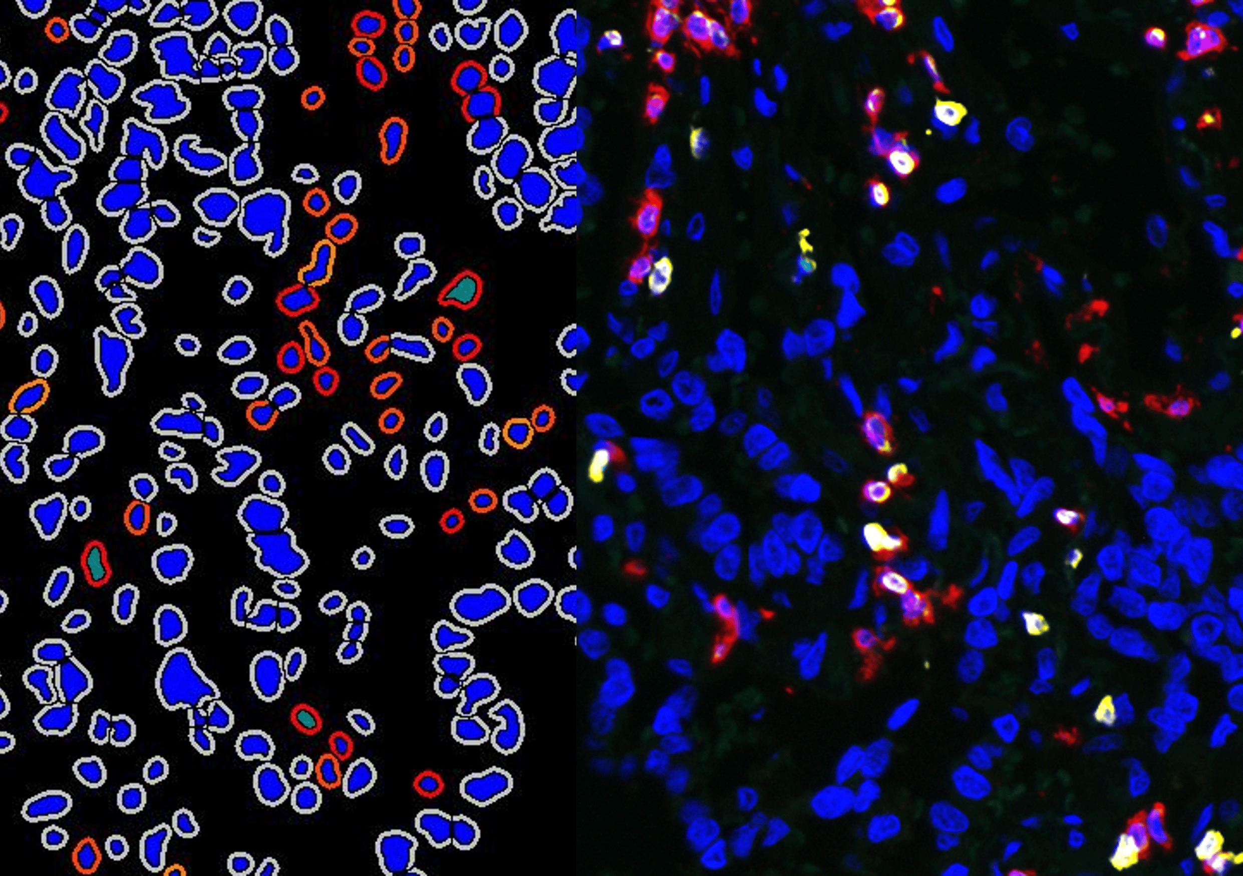
- The PhenoImager© solution from Akoya allows fast scanning of samples stained with up to 8 different biomarkers in immunofluorescence.
- Cerba Research laboratories are set up to provide remote reading solutions to our customers.
- A fast, secure and validated solution is used for the transferring of large image files.
- We offer remote viewing of images hosted on our own servers to avoid unnecessary downloading.
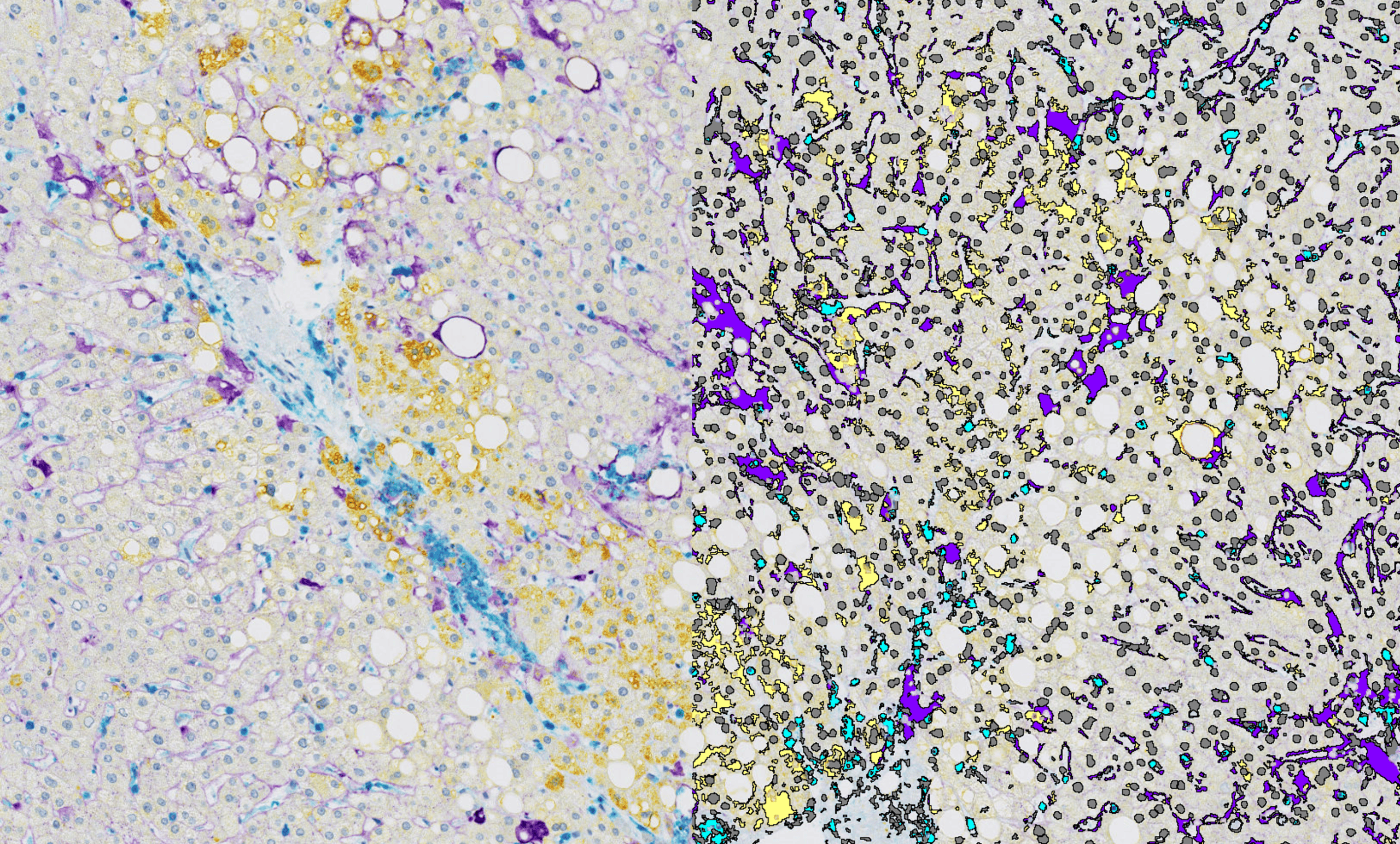
- Getting the best data from stained histological sections cannot always rely on pathologist readings, particularly when multiplexing solutions are required. Our image analysis team is dedicated to providing custom and flexible solutions to extract quantifiable data from biopsy images since 2014. They will suggest expert approaches to get the best dataset from your samples.
- Our multiplexing capabilities allows for accurate phenotyping of cell subpopulations based on the expression or absence of expression of combinations of biomarkers, just as with flow cytometry.
- Moreover, additional data on the spatial organization of the tissue can be obtained, providing information on cell interactions that are critical, especially in immuno-oncology.
We Use The Most Trusted Image Analysis Software
Cerba Research uses the most widely adopted image analysis solutions to allow for easy transfer between our labs and our customers, and make it easier to perform both quality controls and additional analysis on the images we have generated. Having access to multiple softwares also provides extended capabilities to offer the most relevant and efficient method to generate data.
Visopharm® Software
Visiopharm® is used for custom analysis solutions, where the analysis of combinations of features can be automated to a high-level of complexity.
Halo® Software
Halo® (Indica Labs) is an open solution with off-the-shelf modules for quickly actionable and high-throughput solutions for tissue or micro-arrays analysis.
Automated Processing Of Adjudication Scenarios
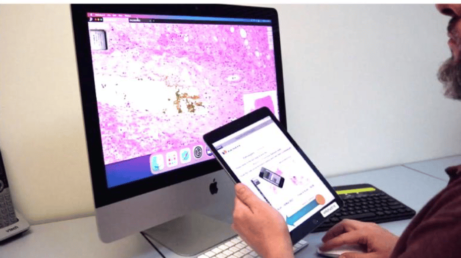
Cerba Research has designed a proprietary solution for automated adjudication and short turnaround time in clinical trials where regulatory authorities request multiple readers to analyze each biopsy.
Each pathologist is equipped with a dedicated tablet to scan the sample ID inserted into the biopsy image and report on the results directly. Scores are compared by an algorithm validated specifically for each trial/scenario, triggering the request for an additional reader when necessary. The single final consensus result is then reported in the clinical trial database.
This solution offers unequaled speed and reliability when multiple readings are required in a short time frame, like, for example, biopsies for the inclusion of patients in NASH trials.
Reach out to our experts and see how we can help advance your clinical trial with digital imaging
Contact Us