Maximize the Protein Data From Your Tissue Samples
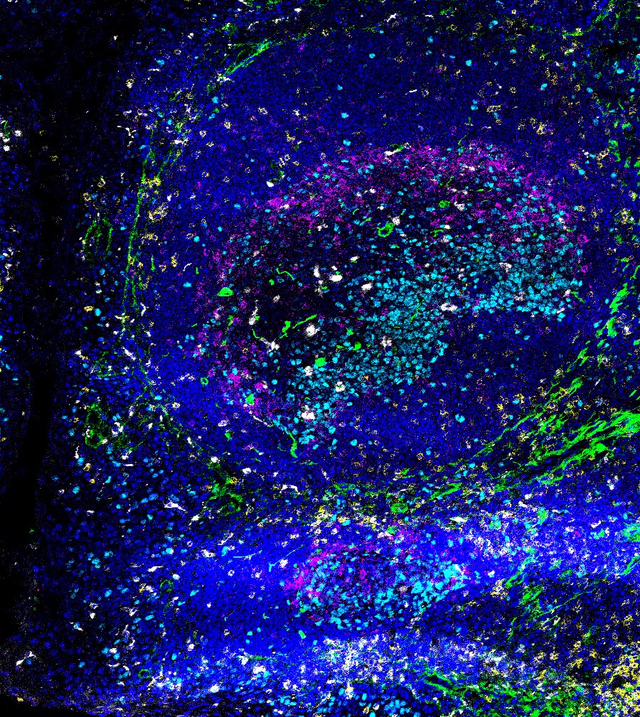
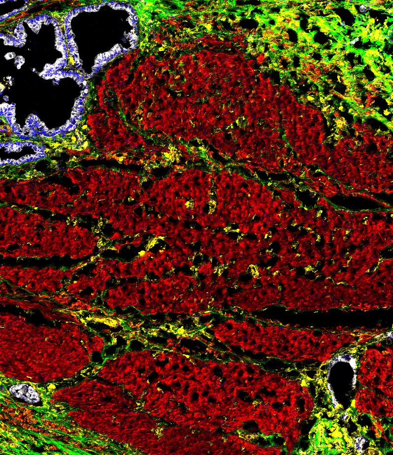
Imaging mass cytometry (IMC) combines antibody-based detection of proteins on tissue with CytTOF® detection. Thanks to the measurement by mass of metal tags cleaved from the antibodies, imaging mass cytometry completely eliminates limitations related to autofluorescence and overlap of emission spectra that come with IF multiplex IHC. This allows for the analysis of up to 37 protein biomarkers per panel.
Cerba Research uses the Hyperion® Imaging System (Standard Biotools Inc.) to analyze and image high numbers of protein biomarkers at the single-cell level. Thanks to this high-resolution spatial information, the platform provides unprecedented value for discovery programs aimed at understanding cellular interactions and biomarker expression profiles at the tissue level.
Our tissue analysis team has experience utilizing the standard Maxpar immuno-oncology panels from Standard Biotools. We also provide a standard workflow to evaluate the performance of your preferred IHC antibody in the IMC format and integrate it in a standard panel.
Various options are available to analyze the data obtained from imaging mass cytometry (IMC) experiments. Our image analysis team will be able to suggest the best approach for your project at the design stage and provide you with high-relevance data from the validation of the solution to the final report.

Reach out to our experts and discover how we can advance your research with Imaging Mass Cytometry
Contact Us